Citation: Seyfang K, “Powder Microdosing with 100% In-Line Fill Weight Control by X-Ray”. ONdrugDelivery Magazine, Issue 85 (Apr 2018), pp 44-47.
Karlheinz Seyfang discusses the need for filling verification in pre-metered DPI blister filling lines, going on to detail the role X-ray analysis could play in an automated verification process.
INTRODUCTION
The Global Asthma Network estimates that approximately 340 million people worldwide suffer from asthma.1 Every day, almost 1000 asthma sufferers die from this disease.2 In 2015, chronic obstructive pulmonary disease (COPD) caused about 3 million deaths worldwide and is forecast to become the third most common cause of death in the next few years.3 Pulmonary administration is the preferred treatment method for these respiratory diseases, in particular pressurised metered dose inhalers (pMDIs) and dry powder inhalers (DPIs). These devices enable the targeted application of locally active drugs, such as anti-inflammatory corticosteroids or bronchodilators.
“Whenever possible, a system for the 100% in-line verification of fill weights should be integrated when processing inhalation powders…”
Due to the large inner surface of the lung and the thin epithelium layer between alveoli and blood vessels, pulmonary administration also enables the use of systemically acting drug substances. In this capacity, characterised by a rapid onset of effect (such as with opiates) and by the potential to administer relatively large molecules (such as insulin), inhalation offers an attractive alternative to oral or invasive parenteral routes of drug delivery.
DPIs have superior user-handling characteristics and drug substance stability compared with other delivery systems. Within the sphere of DPI design, preference is given to devices that utilise individually sealed powder portions that have been pre-metered during production, due to their positive effect on dosing accuracy during application. The active pharmaceutical ingredient (API) particles must have an aerodynamic diameter no greater than approximately 5 μm to penetrate into the deep lung. Powders containing a high proportion of such particles are generally very cohesive, have poor flow properties and exhibit a tendency to adhere to surfaces. Together with the low fill weight, these properties lead to an increased risk of under-dosing occurring in some individual units during production.
Transient process deviations like this could be caused by, for example, bridging of the powder in the feeding system. Issues of this kind cannot be detected with sufficient reliability by statistical in-process controls. Therefore, whenever possible, a system for the 100% in-line verification of fill weights should be integrated when processing inhalation powders. Such a system must be capable of capturing and evaluating the data in real time. However, it does not need to be as accurate as a weighing cell since the focus is on the detection of outliers and their reliable rejection.
BLISTERS FOR DPIS
Ideally, DPIs with pre-metered powder doses should contain a month’s supply, usually 60 doses, in a minimum amount of space. This has an impact on DPI design and therefore on fill technology. In the case of blister-based DPIs, the blister cavities are designed to be as small as possible and, in order to make optimum use of the volume, are usually filled to the brim. Special fill methods are required for production since conventional dosing methods, such as dosator or vacuum dosing drum, cannot typically achieve a 100% filling level.
The so-called membrane filler is a proprietary technology, with which even small target receptacles can be filled completely.4 The system is operated on special thermoforming lines and enables the simultaneous filling of up to 80 blister cavities per machine cycle. A welcome side-effect during dosing is the masking of the sealing surface with the blister web, avoiding contamination by powder particles and thus the occurrence of poorly sealed blister strips. Figure 1 shows the operating principle of the membrane filler.

Figure 1: Operating principle of membrane filler for filling of blister cavities up to the brim.
The blister cavity (6) to be filled is covered with an air-permeable filter membrane (5) connected to a pressure/vacuum system (3). An elastic base (4) provides the air-tight seal. A capillary (2), establishing the connection between blister cavity and powder reservoir (1), ends in the membrane. The dimensions of the capillary are so small that the inhalation powder cannot flow freely purely by gravity. However, if a vacuum pulse is generated through the membrane inside the blister cavity, the powder will flow into the cavity until it is filled to capacity.
Since the fill head of the membrane filler must rest on the blister to be filled, the integration of a capacitive method already in use for the in-line control of fill quantities5 was not possible. An alternative method for the control of the filled-to-capacity blister cavities had to be found.
SELECTION OF A SUITABLE CONTROL TECHNOLOGY
The optical control of filling material in blister packages has long been state of the art. Today’s camera systems are not only capable of detecting missing objects, but also damage, colour and shape deviations, and double filling. This also works with non-translucent aluminium laminates, which are used for most DPI blisters because they provide the necessary moisture barrier. However, it has been found that only those under-filled cavities which are well below 75% of the target fill quantity are detected. In another approach, the topography of the powder surfaces was determined by means of a 3D laser scanner in order to calculate the dosed volume in the cavities based on the theoretical blister geometry. Unfortunately, inhomogeneities and small voids inside the powder-fill such as are prone to occur with such cohesive materials, are not detectable with this method.
Alternatively, a fill-quantity control method using X-ray technology can be utilised, as it is able to “screen” the closed blister. The absorption of the X-rays by the respective material essentially depends on its mass and is independent of the actual volume of the individual blister chambers. Furthermore, the blister webs can also be inspected after sealing, making it easy to integrate such a system into the filling machine and greatly simplifying the handling of rejected blisters filled with highly potent drug substances.
“The evaluation of the detector images resulted in a greyscale value for each blister cavity, which could subsequently be compared with the actual fill quantity…”
Finally, it is safe to assume that the powder will not change during X-ray analysis as the product is exposed to a maximum irradiation dose of 120 μSv, roughly equivalent to the radiation exposure of a passenger during a flight from Frankfurt to New York and back. Uehara et al recently demonstrated that active ingredients in tablets do not change in their quality after being exposed to an X-ray dose of 300 Gy, which is 2.5 million times higher.6
X-RAY CONTROL – FEASIBILITY
X-ray systems are widely used for process monitoring in food packaging in order to detect damage to containers, underfilling and impurities, or foreign bodies. Until now, X-ray systems in pharmaceutical production and packaging have been something of a rarity. Here, too, they are primarily used for the detection of foreign particles, primary packaging defects and assembly faults. The quantitative determination of fill quantities using X-ray technology remains unusual. Furthermore, when it comes to powder-filled blisters, the relatively small powder dose is usually packaged into aluminium laminates. The absorption of X-rays, however, not only depends on wavelength, density and thickness of the irradiated material, but also strongly depends on its atomic number. The 3–4 mg of aluminium contained in the barrier layer per blister cavity, with an atomic number of 13, is thus a high background against the powder (primarily carbon, atomic number 6).

Figure 2: Left: Test setup for feasibility study: 1) X-ray source; 2) Camera; 3) Blister web; 4) Hexapod. (source: Xicron GmbH). Right: Blister web pockets with different fill levels.
For a first study, six-lane blister strips were manufactured with 60 cavities per lane. Some were filled correctly (13 mg), others had several cavities that were manually half emptied or vacuumed out completely (Figure 2). The measurements were carried out with different settings for tube voltage and/or cathode current in order to estimate the achievable measurement accuracy. In order to largely compensate for the influence of material tolerances, i.e. thickness of the carrier strip, as well as the intensity fluctuations of the X-ray tube and the detector on the measurement result, the area encircling the blister cavities was included in the calculation of the effective fill quantity as a kind of “tare weight”.
The evaluation of the detector images resulted in a greyscale value for each blister cavity, which could subsequently be compared with the actual fill quantity. The fill quantity was weighed after determining the gross/tare weights of the cut out individual blister cavities (Mettler MX5, resolution 1 μg). The measured greyscale values correlated quite well with the fill weights (R²=0.968, n=360). One mg of powder corresponded to about 20 greyscale values. Consequently, a sufficiently high resolution was expected in order to detect significantly underfilled blister cavities.
X-RAY CONTROL – INTEGRATION
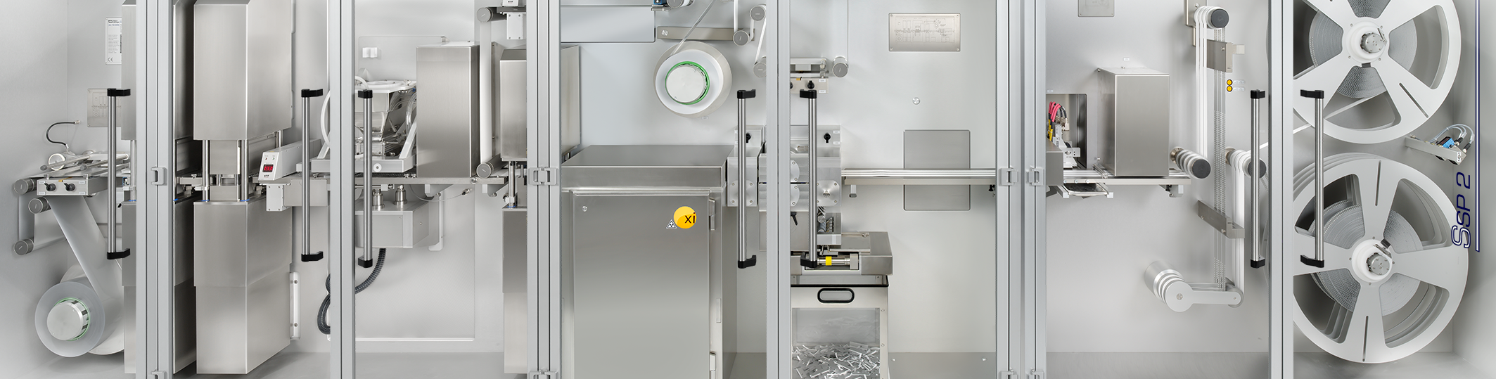
Following the encouraging results of the preliminary study, a production system was designed which can be operated automatically and integrated into existing intermittent blister lines. The size of the flat panel detector was chosen so that up to eight lanes per cycle (this means 8 x 10 blister cavities) could be simultaneously controlled. The module was integrated into the machine after the sealing station (Figure 3). The sealed blisters are X-rayed, then pass through the longitudinal cutting station where they are cut into three or four double strips. This is followed by printing the variable data before the strips are wound up for intermediate storage. Based on the printed data, the winding and assembly machine later detects any non-conforming single blister strips and discharges them from the assembly process.

Figure 3: Machine for the manufacture of powder-filled blister strips: 1) Forming station; 2) Membrane filler; 3) Sealing station; 4) IPC X-ray module; 5) Longitudinal cutting; 6) Printing with camera control; 7) Winding.
The process checks blister cavities when the blister web is stationary for up to one second. During this period, several camera images are generated which are then used to calculate an average greyscale value for the evaluation of the fill quantity. The detector used has a pixel size of 50 μm and the X-ray tube generates a maximum output of 105 kV/450 μA (approximately 50 W). An internal compensation is carried out automatically in order to calibrate the system. Then some empty blisters are measured that serve to detect the impact of deformed and non-deformed blister foil on the greyscale values, as well as the impact caused by their position in the image window. Subsequently, filled blisters were measured, recovered after the longitudinal cutting station and weighed with an automatic checkweigher.
In order to test the system, blisters were filled with lactose monohydrate (Lactohale LH200, DFE Pharma, Goch, Germany). Figure 4 shows the evaluation of a test batch in which 48 blister samples with 60 cavities each (a total of 2880 blister cavities) were weighed. In order to simulate possible under-filled cavities, individual fill nozzles or partial surfaces of the filter membrane were blocked so that some markedly under-filled blister cavities occurred. The deviation of the fill quantity prediction, based on the greyscale values of the checkweigher data, was in the range of -0.85 mg and +0.73 mg, respectively, corresponding to a standard deviation of 0.24 mg. With a target fill weight of 13 mg, this corresponds to a relative standard deviation of 1.8%. The method is sufficiently accurate throughout the entire measurement range to detect individual blisters outside specified limits with a maximum deviation of +5–6%.

Figure 4: Correlation between fill quantities and the calculated values (48 X 60 blister cavities).
We also examined active ingredient containing powder mixtures with a typical combination of beta agonist and corticosteroid. As to be expected from their atomic composition, they showed the same absorption behaviour.
ACKNOWLEDGEMENT
The author would particularly like to thank Mr Jan Rimbach, Xicron GmbH, and Mr Joachim Fahrian, Harro Höfliger Verpackungsmaschinen GmbH, for their support in the compilation of this publication.
REFERENCES
- “The Global Asthma Report 2014”. Global Asthma Network, 2014
- Global Strategy for Asthma Management and Prevention Online Appendix. Global Initiative for Asthma, 2017. (goldcopd.org/download/390/ Accessed March 2018)
- “Chronic obstructive pulmonary disease (COPD)”. WHO Fact Sheet, November 2016. (www.who.int/mediacentre/factsheets/fs315/en/ Accessed March 2018)
- Weigel M, “Fülleinrichtung zum volumetrischen Dosieren von Pulver”. Patent EP2195244 B1, 2012.
- Borchert S, Bullinger T, Seyfang K, “100%-Kontrolle – ohne Leistungsverlust”. TechnoPharm, 2015, Vol 5(5), pp 280–284.
- Uehara K et al, “Effect of X-ray exposure on the pharmaceutical quality of drug tablets using X-ray inspection equipment”. Drug Dev Ind Pharm, Jun 2015, Vol 41(6), pp 953–958.