

Citation: Vasilakos J, “Microneedles for Intradermal Delivery of Cancer Vaccines”. ONdrugDelivery, Issue 116 (Jan 2021), pp 5–9.
John Vasilakos discusses the immuno-oncology market. He examines the role of cancer vaccines in supplementing other immuno-oncology therapies, as well as the value of delivering cancer vaccines intradermally.
Throughout 2020, the pharmaceutical industry received ample public scrutiny around its search for covid-19 vaccines and therapeutics. However, even with so much focus rightfully centred on the race to develop therapies that address the pandemic, covid has not diminished oncology drugs’ colossal importance in the industry. This importance can be measured both in research and development spend and in drug sales. Clinical development spend on oncology therapies in the US is estimated to be more than US$80 billion (£59 billion), which is more than a third of total pharma development spend.1 Meanwhile, drug sales for oncology are expected to exceed $300 billion by 2026, representing roughly a fifth of the total pharma market.1
“A series of studies suggests that patients treated with checkpoint inhibitors are more than twice as likely to experience a durable response.”
The sustained investment and opportunity in oncology matters because cancer remains a public health crisis. Existing drugs and treatment regimens still have many gaps and much room for improvement. These gaps should be addressed by not only optimising the drugs that are used to treat cancer, but also optimising the devices that deliver these drugs.
This article focuses on the sizable immuno-oncology subsegment, which is expected to grow to nearly $100 billion by 2026 – growth of over 20% per annum.1 It discusses a dominant immuno-oncology drug class (checkpoint inhibitors) and their shortcomings, considers the role of cancer vaccines in supplementing other immuno-oncology therapies and the value of delivering cancer vaccines intradermally. Kindeva Drug Delivery’s microneedle-based drug delivery platform will be used as an example of how to address many of the industry’s unmet needs with respect to intradermal delivery.
THE CURRENT LANDSCAPE OF IMMUNO-ONCOLOGY THERAPEUTICS
“The number of T-cell targeted immunomodulators in the pipeline has more than doubled since 2017, with over 200 programmes targeting either PD-1 or PD-L1.”
In the last decade, immune checkpoint inhibitors, such as pembrolizumab (Keytruda, PD-1 antagonist) and ipilimumab (Yervoy, CTLA-4 antagonist), have emerged as a dominant class of immuno-oncology therapeutics. There is a strong case to be made in favour of checkpoint inhibitors based on the durability of the patient response. In essence, this means that a patient can meaningfully extend their life expectancy when these drugs work.
A series of studies suggests that patients treated with checkpoint inhibitors are more than twice as likely to experience a durable response – and roughly 30% of these patients experienced overall survival (OS) that was more than twice the patient population’s median OS.2 Moreover, combination therapies, in which the patient is treated with two or more checkpoint inhibitors, have had an even more promising impact on patient survival.3
Among the most prevalent of the checkpoint inhibitors are programmed cell death protein 1 (PD-1) and programmed cell death ligand 1 (PD-L1) inhibitors. PD-1 is a T-cell receptor that helps downregulate or inhibit immune responses. In the normal course of events, PD-1 engages with PD-L1 on antigen-presenting cells – resulting in the inhibition of T-cell function, such as cytotoxicity. Therefore, PD-1 functions as a brake on T-cell activity, thereby preventing over activation of the immune system, which could result in deleterious effects on healthy tissues. Cancer has found a way to take advantage of this normal regulatory system. Cancer cells can produce PD-L1, which downregulates T-cell function following PD-1 engagement. Therefore, tumour cells that express the ligand PD-L1 block the tumour-killing function of T-cells by engaging T-cell PD-1.

Figure 1: Number of T-cell targeted immunomodulators and cancer vaccines in development globally.
As evidence for their durability grows, checkpoint inhibitors have been commercially successful for many types of cancer, and their popularity in clinical trials has continued to grow. Specifically, the number of T-cell targeted immunomodulators in the pipeline has more than doubled since 2017 (Figure 1), with over 200 programmes targeting either PD-1 or PD-L1 (Figure 2).4 However, despite the promise, there is still a clear need for improvement. The approximately 30% of patients who experience higher OS represent a significant achievement, but approximately 70% of patients require additional therapy.5 Indeed, combination therapy has become the norm for oncology patients.

Figure 2: Number of immunomodulators targeting PD-1/PD-L1 in development globally.
Moreover, checkpoint blockade therapies have recently been approved for use for several cancers. For example, the US FDA has approved Keytruda (Merck, Kenilworth, NJ, US) for use with chemotherapy (carboplatin) to treat metastatic head and neck cancer. In addition to chemotherapeutics, other combination strategies are being evaluated with checkpoint blockade therapies, such as kinase inhibitors, anti-angiogenics, immune-stimulating cytokines and multiple combinations of checkpoint blockade therapies and vaccines.
THE ROLE OF CANCER VACCINES AND COMBINATION THERAPY WITH CHECKPOINT BLOCKADE
Within the immuno-oncology space, there is lots of energy around cancer vaccines. There are an estimated 840 cancer vaccines currently in development (Figure 1),4 with up to 600 companies developing them. In most situations, these vaccines are being developed as combination therapies, intended to be delivered to patients in conjunction with checkpoint inhibitors.
“The question of how and where cancer vaccines are delivered to the body should not be overlooked. The route of administration may impact the ultimate success rates of these vaccines.”
Cancer vaccines are compelling because checkpoint blockade therapies are not successful at eliminating all tumours in all patients, especially when used in isolation. These immunotherapeutics are most effective when the tumour microenvironment (TME) contains tumour-infiltrating T-cells (TILs).6 The TME can be characterised as cold (non-T-cell inflamed) or hot (T-cell inflamed). Hot tumours are characterised by T-cell infiltration and molecular signatures associated with immune activation, whereas cold tumours exhibit T-cell absence or exclusion. In general, hot tumours present higher response rates to checkpoint blockade therapies. Therefore, various efforts have focused on converting non-inflamed cold tumours into hot tumours to achieve a better clinical response.
To solve the problem of converting cold tumours to hot ones, numerous strategies have been employed, such as targeting tumours with immune stimulators or inhibitors of immune suppressor cells. Another approach currently being evaluated is cancer vaccination in combination with checkpoint blockade. Vaccination makes sense in the context where the patient exhibits minimal cancer-specific T-cell immunity. The idea is to increase the number of tumour-specific T-cells with the vaccine and prevent or inhibit their inactivation, or enhance their proliferation, using checkpoint blockade therapies. In other words, cancer vaccines can induce tumour-specific T-cells in those patients that lack anti-tumour T-cells.
Some of the earliest cancer vaccines available on the market proved to be only marginally effective. Now, the industry has become much wiser about cancer vaccines with respect to antigen selection, patient selection and combination therapies to use with vaccines, which adds to their promise. Regarding patient selection or targeting, a better understanding of the patient’s tumour burden and immune status help define which patients may benefit from vaccination. Conducting vaccine trials only on patients with late-stage cancer is not ideal, because those patients often have severely compromised immune systems. For cancer vaccines to work, the patient needs to have a functional immune system. Therefore, cancer vaccines should be delivered to and tested on patients in earlier stages of the disease.
Other improvements in the development of cancer vaccines have resulted from the use of more optimal adjuvants or viral vectors that enhance cytotoxic T-cells and interferon-gamma producing T-helper cells, and the use of cancer antigens that are more commonly expressed on multiple tumour types or antigens that are unique to the patient (neoantigens).
Continued innovation in cancer vaccines and their formulations is essential – but the question of how and where cancer vaccines are delivered to the body should not be overlooked. The route of administration may impact the ultimate success rates of these vaccines.
INTRADERMAL DRUG DELIVERY OF CANCER VACCINES
As previously discussed, cancer vaccines induce tumour-specific immunity. In an ideal world, cancer vaccines would be delivered directly into immune organs such as the lymph nodes, the spleen or the skin itself. When delivered subcutaneously or intramuscularly, cancer vaccines are deposited into parts of the body where immune cells do not normally reside. By contrast, intradermal delivery would deliver the vaccine directly into the dermis, where immune cells do reside. There is evidence that demonstrates a comparative advantage of intradermal delivery over intramuscular delivery.7 Of salience to cancer vaccines, intradermal vaccinations have led to enhanced immune responses in many cases.8 While intradermal delivery of cancer vaccines seems ideal, its biggest limitation is a practical one: it is difficult to achieve precise and reproducible delivery to the intradermal layer using traditional delivery devices. One challenge is that the dermis is thin – approximately 1,800 μm in depth. To achieve this, the delivery device would need to deliver the vaccine to a depth of 500–1,500 μm and do so reliably. The lack of a reliable delivery method has limited the use of intradermal vaccination.9

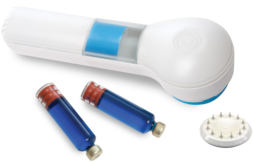
Figure 3: Kindeva’s hollow microstructured transdermal system (hMTS).
Kindeva has advanced the development of delivery devices that help overcome the challenge of intradermal delivery. With microneedles, Kindeva’s hollow microstructured transdermal system (hMTS) facilitates the reliable and reproducible delivery to the dermis. Kindeva’s hMTS device is an injector that includes a drug cartridge to contain the liquid formulation and an array comprised of 12 hollow microneedles (Figure 3). The cartridge can be loaded up-front or at the time of use, depending on the drug’s requirement. At the time of use, the applicator delivers the microneedle array into the skin; the liquid formulation will then move through the hollow microneedles and deliver the drug into the dermis. The drug delivery system is intended for the intradermal space, which makes the injection time longer than is typically experienced with standard vaccine injections. Depending on the formulation, the patient and the delivery site, total hMTS injection time is typically less than two minutes per mL.

Figure 4: Anatomy of the skin.
The two main features that make Kindeva’s hMTS platform well suited to deliver cancer vaccines are the depth of delivery and the volume of delivery. For depth, Kindeva has refined the device design to achieve reproducible intradermal delivery, using microneedles with a length of either 1,000 μm or 1,500 μm. Cancer vaccines administered via hMTS will be delivered to a shallower part of the skin compared with vaccines delivered subcutaneously (Figure 4), thus delivering the vaccine to the immune-cell-rich dermis and elicit a more robust immune response.
In terms of volume, hMTS can deliver up to 2 mL. This is a meaningful increase in capacity compared with other intradermal and non-intradermal delivery devices currently used to deliver vaccines. While the hMTS device is currently in development, it is already well positioned to be the device-of-choice for biopharma companies developing cancer vaccines. Kindeva’s device has been used in partners’ Phase I and IIa drug clinical studies in the US,10-13 and the development of a device suitable for Phase III/commercial use is progressing. Moreover, Kindeva’s manufacturing capabilities are in place to meet preclinical and clinical needs.
CONCLUSION
Immuno-oncology therapies, such as checkpoint inhibitors, are designed to facilitate the ability of the patient’s immune system to effectively attack and kill tumours. Over the last decade, there has been demonstrable success, with blockbuster therapies achieving significant increases in patient survival rates. Cancer vaccines can be used in combination with these checkpoint inhibitors to make cold tumours hot, thereby increasing the overall success rate. New oncology drug classes continue to crop up and gain momentum, which provides a reason for optimism.
However, innovation in the pharmaceutical industry is not exclusive to the discovery of novel drugs and vaccines. Innovation in drug delivery devices can also play a meaningful role in improving how the medical industry treats cancer. For example, devices that enable reproducible intradermal delivery should be seriously considered by biopharma companies developing cancer vaccines. Devices like Kindeva’s hMTS platform are capable of delivering cancer vaccines directly to the dermis, with its relative abundance of immune cells, increasing the potential that the drug is effective in improving patient survival rates.
REFERENCES
- “World Preview 2020, Outlook to 2026.” EvaluatePharma, 2020.
- Pons-Tostivint E et al, “Comparative Analysis of Durable Responses on Immune Checkpoint Inhibitors Versus Other Systemic Therapies: A Pooled Analysis of Phase III Trials.” JCO Precision Oncology, 2019, Vol 3, pp 1–10.
- Larkin J et al, “Five-Year Survival with Combined Nivolumab and Ipilimumab in Advanced Melanoma.” N Engl J Med, 2019, Vol 381, pp 1535–1546.
- Upadhaya S, Hubbard-Lucey VM, Yu JX, “Immuno-oncology drug development forges on despite COVID-19.” Nat Rev Drug Discov, 2020, Vol 19, pp 751–752.
- Haslam A, Prasad V, “Estimation of the Percentage of US Patients with Cancer Who Are Eligible for and Respond to Checkpoint Inhibitor Immunotherapy Drugs.” JAMA Netw Open, 2019, Vol 2(5), e192535.
- Duan Q et al, “Turning cold into hot: firing up the tumor.” Trends in Cancer, 2020, Vol 6(7), pp 605–618.
- Teunissen MB, Haniffa M et al, “Insight into the immunobiology of human skin and functional specialization of skin dendritic cell subsets to innovate intradermal vaccination design.” Curr Top Microbiol Immunol, 2012, Vol 351, pp 25–76.
- Ogai N, Nonaka I et al, “Enhanced immunity in intradermal vaccination by novel hollow microneedles.” Skin Res Technol, 2018, Vol 24(4) pp 630–635.
- Kim YC, Jarrahian C et al, “Delivery systems for intradermal vaccination.” Curr Top Microbiol Immunol, 2012, Vol 351, pp 77–112.
- Gramza A et al, “Phase 1/2 Open- Label, Multi-Center Trial of SNS-301 Added to Pembrolizumab in Patients with Advanced Squamous Cell Carcinoma of the Head & Neck.” Poster Presentation, 2020 SITC Annual Meeting.
- Algazi A et al, “Early Safety and Efficacy of a Phase 1/2 Open-Label, Multi-Center Trial of SNS-301 Added to Pembrolizumab in Patients with ASPH+ Locally Advanced Unresectable or Metastatic/Recurrent Squamous Cell Carcinoma of the Head and Neck.” Poster Presentation, 2020 EMSO Annual Meeting.
- Block MS et al, “Th17-inducing autologous dendritic cell vaccination promotes antigen-specific cellular and humoral immunity in ovarian cancer patients.” Nature Communications, 2020, Vol 11(1), Article 5173.
- Gustafson MP et al, “Preliminary manufacturing, safety, and immune monitoring results of an allogeneic tumor lysate-pulsed dendritic cell vaccine for patients with newly diagnosed glioblastoma.” Poster Presentation, 2016 SITC Annual Meeting.