
Citation: Seaman P, Palmer D, “Opsisporin: A Long-Acting Drug Delivery Approach for Uveitis”. ONdrugDelivery Magazine, Issue 63 (Jan 2016), pp 32-35.
Here, Paul Seaman and Daniel Palmer discuss Midatech Pharma’s next generation treatments for auto-immune ocular diseases, which use a combination of innovative, sustained-release drug products.
Current treatments for eye diseases often do not achieve the required level of effect. Topical therapies have low efficacy and bioavailability as a result of factors such as anatomical and physiological barriers, tear formation, and lymphatic drug clearance. Systemic therapies have high incidence of adverse effects, poor tolerability, and limited efficacy. Frequently, patients are non-compliant due to unpleasantness, pain, or inconvenience of the treatment options.
Midatech’s sustained-release technology, delivered directly to a specific ocular space, addresses these challenges. Localised long-acting delivery of API offers marked advantages and consistency over oral administration since drugs are delivered to the target site in therapeutic concentrations, off-target side effects of high-dose systemic delivery are avoided, and patient experience is improved via minimally-invasive, convenient and infrequent administration.
TECHNOLOGY
Our proprietary microsphere engineering platform can use a wide range of biomaterials to encapsulate ocular drug candidates into micron sized particles (of diameter ~25μm). These include glucocorticoid anti-inflammatories, anti-VEGFs and immunosuppressants for diseases such as glaucoma, AMD and non-infective uveitis. Long-acting treatment is achieved using formulations of biodegradable polymers (including polylactides) to control the release of API over a period of 3-6 months following a single intravitreal or subconjunctival injection. Monodisperse microspheres may be readily injected via minimally-invasive needles as fine as 30G.
In formulating small molecules, biopharmaceuticals and PEGylated species, Midatech focuses on developing products that provide high drug loading, with minimal initial burst release. These characteristics are essential to the development of safe and effective ophthalmic therapies. The formulation tuning and process control to achieve this requires precision know-how and novel technology to ensure control over particle size, morphology and drug kinetics.
Our technology represents a step-change in control for microsphere manufacturing, enabling emulsion-free synthesis with both product monodispersity and processing efficiency built-in. Further, the encapsulation process is engineered to avoid the use of poorly-tolerated organic solvents such as dichloromethane. The manufacturing system has been designed for total compatibility with the aseptic production environment and standard isolator technology, and is proven for sterile synthesis and filling of clinical trial materials.
This level of flexibility enables Midatech to formulate products to provide release profiles and dosage regimes that treat ocular disease states in the most efficacious manner.
UVEITIS
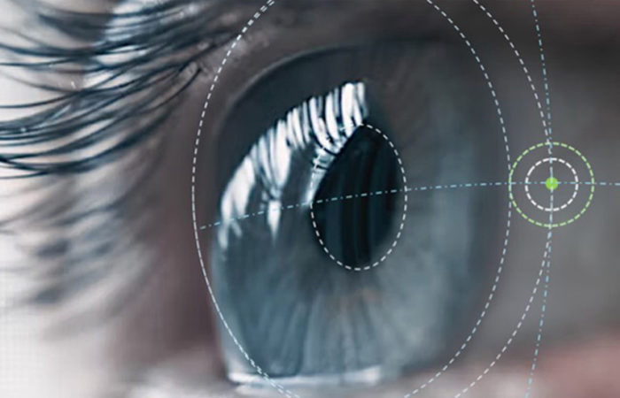
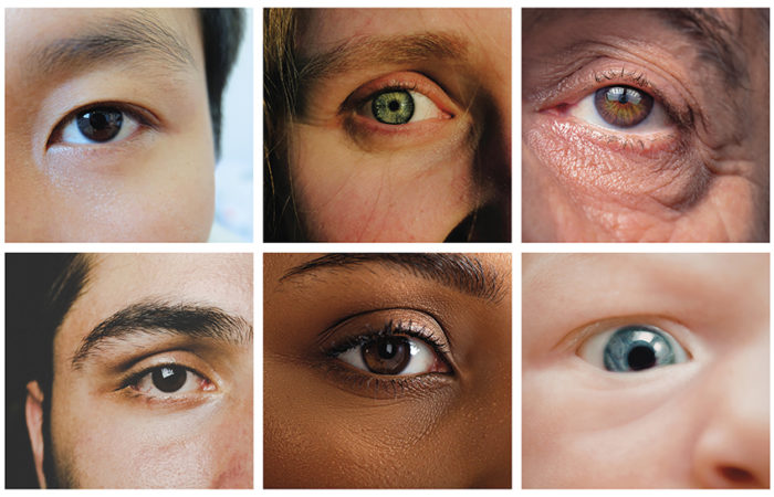
One such application of the Midatech technology is for the treatment of posterior uveitis, an intra-ocular inflammatory condition affecting one or more parts of the uvea which includes the iris, the ciliary body, and choroid of the eye, although other ocular structures such as the retina and the vitreous body may also be secondarily involved. Aetiology is varied and includes infectious causes (e.g. tuberculosis, toxocara) and non-infectious pathogenesis. Non-infectious causes include systemic auto-immune disorders (e.g. rheumatoid arthritis, sarcoidosis, Lupus, Behçet’s disease), trauma, and rarely can also be drug induced. However, many cases of uveitis are idiopathic in nature. Uveitis is a major cause of significant visual loss in young and middle aged adults in the Western world. The burden, to society and the patient, resulting from uncontrolled uveitis is significant.
“Depot-based drug delivery is strongly required in ocular disease, due to the burden of frequent intravitreal injections and lack of any satisfactory treatments to date…”
Uveitis is a major cause of new cases of blindness; in the US, it is estimated that uveitis causes an estimated 30,000 new cases of legal blindness and results in 2.8-10% of all blindness annually. Uveitis can be classified on the basis of the aetiological factors (e.g. infectious, non-infectious, drug induced etc), or most commonly, on the anatomical location of the disease, anterior, intermediate, posterior, and panuveitis. Until recently, not all EU countries had uveitis as a listed indication and it is still not an approved indication in the US.
The presenting symptoms tend to vary with the type of uveitis and the severity, and may affect one or both eyes. Anterior uveitis is often accompanied by ocular pain and photophobia. Blurring of vision is less of a characteristic of anterior uveitis, but may be reported in severe cases or by patients with secondary cataracts or macular oedema. Posterior uveitis may be accompanied by some degree of pain and photophobia, but these may not be the predominant symptoms. However, blurred vision is common due to involvement of the macula (inflammation and macular oedema) and opacity of vitreous humour (flare and cell infiltration). Vasculitis and retinal edema are also notable features. It is important to establish the correct diagnosis so that specific treatment to address the underlying aetiology can be initiated, along with treatment to reduce ocular inflammation.
The inflammatory process in the eye(s) must be reduced quickly to avoid long-term consequences. The treatment options for non-infectious uveitis affecting the posterior or intermediate segment of the eye include the use of local and systemic corticosteroids, systemic immunosuppressants, and off-label use of anti-VEGF agents and systemic biologic agents. Patients are routinely initiated on systemic steroids, the mainstay of posterior uveitis therapy, at high doses of up to 1 mg/kg/day and then tapered to ideally less than 5 mg/day. If, after a period of time (ranging from 2-3 months), the steroids cannot be reduced or eliminated, systemic immunosuppressives are started such as cyclosporin, or off-label adalimumab, azathioprine, cyclophosphamide, infliximab, methotrexate, tacrolimus and others. Each of these has the potential for causing serious systemic adverse effects that limit the dose or their use altogether. Intra-ocular administration of sustained release steroids like fluocinolone and dexamethasone are an advance, but risk causing cataracts, glaucoma, and increased intraocular pressure; and the duration of effect seems to be limited and with high relapse rate. The systemic use of cyclosporin for the treatment of non-infectious uveitis of the posterior segment is common practice among uveitis specialists. However, systemic drugs (corticosteroids and immunosuppressive therapy) often fail because of the presence of the blood-retinal barrier that limits drug delivery into uveal tissues.
The important, sometimes life threatening, adverse effects of these drugs limit the maximum dose that can be recommended. The dose given may, therefore, be suboptimal in terms of control of inflammation in the eye even when maximum dosing is prescribed.
In summary, although the treatment options available for patients with non-infectious uveitis of the posterior segment of the eye have recently improved, none is ideal. Topical steroids often do not work and systemic corticosteroid or immunotherapy is limited by the risks of adverse events with protracted use. Ocular inflammation may require the addition of intravitreal steroid injections to prevent permanent damage to vision. These are effective, but have a relatively short duration of action as well as side effects. This medical need has been recognised and implants for the intravitreal administration of steroids have been developed, but again side effects are problematic and efficacy inconsistent. A product that achieves control of inflammation without serious steroid-related side effects would constitute a significant benefit.
OPSISPORIN
OpsiSporin is a treatment under development by Midatech. It comprises encapsulated cyclosporine, a cyclic peptide of 11 residues that has been routinely used as an oral immunosuppressor for organ transplantation since first approval in 1983. The drug’s mode of action is well understood, being the selective inhibition of interleukin-2 release during the activation of T-cells, suppressing cell-mediated immune response. Cyclosporin forms a complex with the cytosolic protein cyclophilin of lymphocytes, especially T cells. This complex inhibits calcineurin, which, under normal circumstances is responsible for activating the transcription of interleukin-2. This results in an immunosuppression that is reversible when treatment is ceased. Therefore, diseases that involve cytokines or immune–related disorders, such as non-infectious uveitis, are potential indications for cyclosporin. Through prevention of T-cell activation and proliferation, cyclosporin has been hypothesized to be a suitable treatment for non-infectious uveitis.
Sustained-release of cyclosporin within the posterior eye is expected to reduce or spare the need for oral drug treatments used in the treatment of uveitis, in particular oral glucocorticosteroids and immunosuppressants; and the literature indicates that the use of intravitreal cyclosporin is effective and well tolerated as described above. Hence, OpsiSporin is expected to be effective in the treatment of uveitis affecting the posterior segment of the eye. In treating the disease and preventing recurrence, its use could remove the need for long-term steroid cover required to treat ocular inflammation avoiding the complications of chronic systemic or intravitreal corticosteroids. Systemic immunosuppression may still be required to address the aetiology of the uveitis, but the dose may be tailored to this objective and not raised excessively in an effort to suppress ocular inflammation with systemic dosing.
Sustained delivery of the cyclosporin is achieved by encapsulation with bioresorbable polymer excipients, such that the endecapeptide is physically entrapped in the polymer matrix. Cyclosporin is released as the poly-ester backbone of the polymer undergoes hydrolysis and is broken in to its constituent monomers, resulting in the gradual erosion of the polymer matrix.
The development of the product targeted a sustained release (SR), microparticle formulation, with the following product attributes:
- Cyclosporin drug loading >15% w/w
- Cyclosporin encapsulation efficiency >80%
- Formation of stable suspension, injectable via ½” 30G needle
- Linear release kinetics maintained for 90-100 days.
Early formulation development focused on identifying parameters for satisfactory drug encapsulation (suitable drug loading and stability) and applying knowledge of SR formulations to produce early formulations aimed at a three-month release duration (established via in vitro release modelling). Formulations have been focused on polymers from the polylactide-co-glycolide family, specifically those containing a high ratio of lactide to glycolide (typically 75-100%).
Additionally, review of the literature indicated that higher-molecular-weight polymers were also well suited to this application. It is known that drug loosely bound to the surface of microspheres can give rise to a rapid drug dissolution profile immediately upon administration; i.e. an undesirable burst release of cyclosporin. Therefore, the manufacturing process involves a post-processing step during which the surface drug is removed and this burst release significantly attenuated. Particle-drug loading was generally very high (~19-21% w/w), and was not significantly affected by particle post-processing.
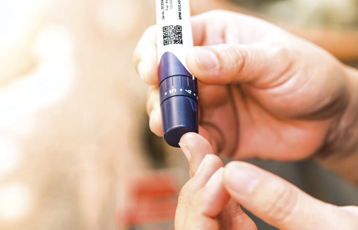
Similar to many microsphere-based SR products, OpsiSporin is presented as a powder that is formed into a suspension prior to dosing. Due to the intravitreal route, where injection volume is limited to 0.1 mL, and the hydrophobic nature of cyclosporine-loaded microspheres, formation of a simply and reproducibly injectable suspension required significant development. Midatech has worked to develop the formulation to make reconstitution rapid, consistent and safe.
The application of microsphere processing expertise has resulted in the introduction of technology that enables OpsiSporin to be reconstituted rapidly with nothing more than water. The formulation excludes novel excipients whilst preventing microsphere aggregation during storage and reconstitution, and maximising suspension stability. As a result, homogeneous suspensions can be formed in under 30 seconds and remain stable for several minutes, meaning preparation in the clinic is minimal and the dosing need not be rushed. Data gathered to date indicate that OpsiSporin is injectable through ½” 30G needles.

Figure 1: Proof of concept in murine model. Increasing TEFI score of retinal imaging indicates worsening of the EAU disease state. Mice receiving intravitreal injection of OpsiSporin (4.5μg cyclosporin) showed a significant reduction in EAU severity compared to those animals that received vehicle alone.
PROOF OF CONCEPT
An in vivo study has illustrated the efficacy of OpsiSporin in the treatment of experimental auto-immune uveitis (EAU) in a murine model. EAU was induced in groups of ten C57/BL6 mice by challenge with retinal antigens interphotoreceptor retinoid binding protein peptide (IRBPp) and Freund’s adjuvant (subcutaneous administration) and pertussis toxin (intraperitoneal administration). Due to the sustainedrelease properties of the OpsiSporin product, intravitreal treatment was administered on a single occasion only (day zero) via 33G needle. A negative control group receiving vehicle only was also injected at day zero. In addition, two groups of ten animals received different doses of oral cyclosporin (in car-boxylmethylcellulose solution), once daily, as positive control treatments.
Increasing TEFI score of retinal imaging indicates worsening of the EAU disease state. Mice receiving intravitreal injection of OpsiSporin (4.5μg cyclosporin) showed a significant reduction in EAU severity compared to those animals that received vehicle alone (Figure 1). Topical endoscopic fundal imaging (TEFI) is a technique used to monitor the progression of retinal degradation in ocular disease. In this study it was used to track the progress of EAU after administration of the IRBPp / pertussis toxin insult. An increasing TEFI score denotes the advancement of the EAU disease state, and hence can be used to observe recovery or reduction in retinal damage and inflammation. Mice receiving intravitreal injection of OpsiSporin (4.5μg cyclosporin) showed a significant reduction in EAU severity compared to those animals that received vehicle alone (p<0.002), supporting the hypothesis that delivery of cyclosporin via the OpsiSporin product has potential for the treatment of non-infectious uveitis in man. A significant reduction in disease development was observed at the later time points, as severity of disease increased in the untreated and negative control groups. Multiple t tests between untreated animals and those receiving cyclosporin either as OpsiSporin or orally showed a significant reduction in disease from day 15.

Figure 2: In vitro testing of OpsiSporin models the release of API from the drug product, with the data plotted as a cumulative mass of drug released. Highly linear release of cyclosporin lasting three months was observed with OpsiSporin. These data show the mass of cyclosporin released per mg of OpsiSporin drug product, and predict a linear release of 0.96 μg/mg/day. Data are the mean of three replicates, and error bars represent standard deviation.
In addition, a significant reduction was observed between those animals receiving intravitreous injection of vehicle with those receiving OpsiSporin containing 4.5 μg cyclosporin. An equivalent reduction in disease development was observed following daily oral administration of 6.7 mg/kg/day cyclosporin, as compared with intravitreous injection of 4.5μg OpsiSporin; efficacy was equivalent whilst total amount of cyclosporine administered was 1000- fold lower with OpsiSporin. The EAU model is self-limiting, and as such is only able to indicate efficacy for up to 28 days.
In vitro modelling of drug release kinetics indicated that the rate of cyclosporin release shown to be efficacious in vivo, were maintained for three months (see Figure 2). It is therefore hypothesised that efficacy will be maintained over the entire target period, facilitating a three-monthly dosing regimen.
CONCLUSION
Depot-based drug delivery is strongly required in ocular disease, due to the burden of frequent intravitreal injections and lack of any satisfactory treatments to date. Solid formulations that are precisely defined, easy to administer into the eye, and able to provide steady drug release over 3-6 months are ideal alternatives for chronic conditions such as non-infective posterior uveitis. In developing its ocular drug delivery platform, Midatech technology may provide a compelling and unique platform to bring significant benefits to patients suffering from uveitis by providing a long-acting, easy-to-use efficacious product that spares the use and thus side effects of treatment with systemic or local steroids, or immunosuppressants.
REFERENCES
- Caspi, Rachel R. “Understanding Autoimmunity in the Eye: From Animal Models to Novel Therapies.” Discov Med, Mar 2014, Vol 17(93), pp 155-162.
- Jabs DA et al, “Guidelines for the Use of Immunosuppressive Drugs in Patients with Ocular Inflammatory Disorders: Recommendations of an Expert Panel.” Am Journal Ophthalmol, Oct 2000, Vol 130(4), pp 492-513.
- Lallemand F et al “Cyclosporine A Delivery to the Eye: A Pharmaceutical Challenge.” Eur J Pharm Biopharm, Nov 2003, Vol 56(3), pp 307-318.
- Pavesio C et al, “Evaluation of an Intravitreal Fluocinolone Acetonide Implant versus Standard Systemic Therapy in Noninfectious Posterior Uveitis.” Ophthalmology, March 2010, Vol 117(3), pp 567-575.
- Thompson, Angus W. “The Influence of Cyclosporin A on T Cell Activation, Cytokine Gene Expression and Cell-mediated Immunity.” Cyclosporin: Mode of Action and Clinical Applications; Springer, Dordrecht, Kluwer Academic, 1989.
- Trusko, B et al, “The Standardization of Uveitis Nomenclature (SUN) Project.” Methods Inf Med, 2013, Vol 52(3), [[ 259-265.
- de Vries J et al, “Cyclosporin in the Treatment of Severe Chronic Idiopathic Uveitis.” Br J Ophthalmol, Jun 1990, 74(6), pp 344-349.